Samson SY WONG
Invasive fungal infections have frequently been caused by opportunistic pathogens in the immunocompromised hosts. Candidaemia commonly occurs in nosocomial bloodstream infections, invasive aspergillosis often complicates neutropaenic patients, and cryptococcal meningitis is a well known AIDS-defining illness. All these opportunistic mycoses can be found worldwide, but penicilliosis is an endemic mycosis that is peculiar to the Southeast Asia and its importance emerged only after the global AIDS epidemic started to take its toll in this part of the world.
Penicilliosis is an infection caused by the fungus Penicillium marneffei (P. marneffei). P. marneffei is a dimorphic fungus, meaning that it may exist in a yeast-like morphology macroscopically and microscopically, as well as a mould form. The transition of dimorphism is dependent on temperature (thermal dimorphism), with the yeast form being produced at 37°C (in host tissues and in culture) and the mould form at 25°C. P. marneffei is the only Penicillium species that exhibits thermal dimorphism and that is regularly pathogenic to humans.
Like many other pathogenic dimorphic fungi, P. marneffei is geographically restricted. Indigenous cases have been reported in most Southeast Asian countries such as Vietnam, Hong Kong, Taiwan, the Philippines, Malaysia, and eastern India, but the largest number of cases occurred in Thailand. In mainland China, cases have been reported in the Guangxi and Guangdong provinces.1,2 The natural reservoir of the fungus and the mode of transmission to humans are unknown. However, it is generally believed that P. marneffei is likely to be present in the soil and humans acquired the infection through inhalation of the fungal spores. The only other animal that has been found to be naturally infected by P. marneffei is the bamboo rat, though this animal is not considered to be a reservoir host of the fungus, nor does it play a role in causing human infections.
Penicilliosis has occurred in immunocompetent hosts and patients with other immunocompromising conditions.3 However, the majority of the cases are seen in patients co-infected with HIV. In Southeast Asia, penicilliosis is considered to be an AIDS-defining illness. In Thailand, for example, penicilliosis is the third commonest opportunistic infection in AIDS patients following tuberculosis and cryptococcal meningitis.4 In Hong Kong, 7.7% of AIDS patients developed penicilliosis during the course of the illness.5
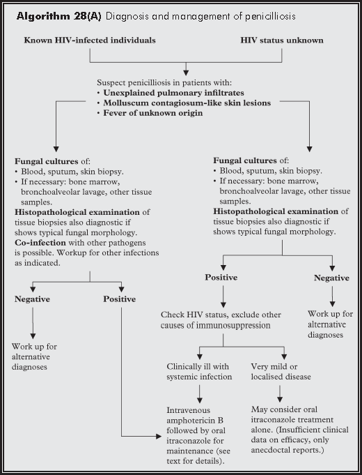
Penicilliosis is a systemic infection resulting in fungaemia, pulmonary infections, skin lesions, and involvement of the reticuloendothelial system. In Hong Kong, patients not infrequently have penicilliosis as the presenting illness that lead to the diagnosis of HIV infection. The initial manifestation is often fever with or without pulmonary infiltrates on the chest radiograph, weight loss, hepatosplenomegaly, lymphadenopathy, and skin lesions. There are no pathognomonic signs on the chest radiograph. Radiographic appearance of the lungs is variable, ranging from diffuse or localised reticular, reticulonodular, alveolar infiltration, cavitary lesion, interstitial pneumonia, lung nodules, and pleural effusion. The presence of skin lesions may sometimes be suggestive of the diagnosis. The most typical lesion is a molluscum contagiosum-like nodule with central umbilication, although other forms of skin lesions (ranging from papules, pustules, ulcers, to subcutaneous abscesses) may be seen as well. Fungaemia occurs in 50%-64% of the patients. Other manifestations include oropharyngeal lesion, intestinal and musculoskeletal involvement. In contrast to cryptococcosis, involvement of the central nervous system in penicilliosis is extremely rare. The CD4 count in HIV-infected penicilliosis patients is almost always less than 100/μL at the time of penicilliosis. Median CD4 counts reported ranged from 5.5/μL (Taiwan), 34/μL (Hong Kong), and 64/μL (Thailand).6,7
As there are no pathognomonic signs and symptoms of penicilliosis, clinical suspicion of the condition is crucial. The presence of fever, pulmonary infiltrates, and the characteristic skin lesions would make the diagnosis very likely. In other situations, relevant laboratory investigations are essential, such as tissue biopsies, bronchoalveolar lavage, and bone marrow cultures. The infection should be suspected in patients with known HIV-infection and presenting with unexplained fever. On the contrary, all patients diagnosed to be having penicilliosis should have the HIV antibody status checked.
The definitive diagnosis of P. marneffei infection still relies on mycological culture. The fungus is readily cultivable in routine fungal cultures from blood, bone marrow aspirate, respiratory tract specimens, and other clinical specimens such as biopsies of skin lesions and fine needle aspiration of lymph nodes. Suspicious lesions should always be biopsied for microbiological and histopathological investigations. The yeast cells in biopsies have a distinctive morphology in tissue sections. They may resemble the tissue phase of another dimorphic fungus Histoplasma capsulatum. However, P. marneffei yeasts divide by fission instead of budding as seen in H. capsulatum. The result is that a transverse septum can be seen in between two P. marneffei cells. The demonstration of thermal dimorphism of the fungus cultures, the presence of a diffusible red pigment in the mould culture, and the typical microscopic morphology of the mould phase all help to confirm the identity of the fungus. Apart from mycological cultures, a number of serological tests have been described. In Hong Kong, an indirect immunofluorescent antibody test is available in some centres.8 The test is valuable in patients with a relatively low fungal load and in whom invasive investigations may not be possible. However, the utility of antibody testing is generally limited by the immune status of the host and in severely immunocompromised patients, a positive antibody test may not always be obtained even in the presence of infection.
Untreated disseminated penicilliosis is almost uniformly fatal. The infection, however, is readily treatable with available antifungals. The most well established regimen consists of intravenous amphotericin B at 0.6 mg/kg/d (others have used doses of amphotericin B from 0.7-1.0 mg/kg/d) for two weeks followed by ten weeks of oral itraconazole at 400 mg/d as maintenance therapy.9 Less severe case may be treated with itraconazole alone. Liposomal amphotericin B has been used successfully for the treatment of penicilliosis in a renal transplant recipient.
Long term secondary prophylaxis with oral itraconazole 200 mg/d is necessary to prevent relapse. In the absence of immuno-reconstitution with HAART, lifelong prophylaxis is probably needed. In patients who are receiving HAART, whether antifungal prophylaxis can be discontinued and when should it be stopped is unknown. However, data from Taiwan showed that secondary antifungal prophylaxis might be discontinued for patients receiving HAART. The median CD4 count at discontinuation was 95/mL and they had received a median of 10.8 months of antifungal therapy and 9.9 months of HAART.7 Newer antifungal agents have not been tested for efficacies against penicilliosis in controlled clinical trials. In vitro studies demonstrated antifungal activities by the newer azoles (e.g. voriconazole and posaconazole), terbinafine, and echinocandins (e.g. anidulafungin and micafungin).10 The newer azoles appears to have very low minimum inhibitory concentrations against P. marneffei.
Wong SS, Yuen KY. Penicilliosis in China. Mycopathologia 2004;158:147-50.
Liyan X, Changming L, Xianyi Z, Luxia W, Suisheng X. Fifteen cases of penicilliosis in Guangdong, China. Mycopathologia 2004;158:151-5.
Wong SS, Wong KH, Hui WT, et al. Differences in clinical and laboratory diagnostic characteristics of penicilliosis marneffei in human immunodeficiency virus (HIV)- and non-HIV-infected patients. J Clin Microbiol 2001;39:4535-40.
Supparatpinyo K, Khamwan C, Baosoung V, Nelson KE, Sirisanthana T. Disseminated Penicillium marneffei infection in southeast Asia. Lancet 1994;344:110-3.
Low K, Lee SS. The pattern of aids reporting and the implications on HIV surveillance. Public Health and Epidemiology Bulletin 2002;11:41-9.
Sirisanthana T, Supparatpinyo K. Epidemiology and management of penicilliosis in human immunodeficiency virus-infected patients. Int J Infect Dis 1998;3:48-53.
Sun HY, Chen MY, Hsiao CF, Hsieh SM, Hung CC, Chang SC. Endemic fungal infections caused by Cryptococcus neoformans and Penicillium marneffei in patients infected with human immunodeficiency virus and treated with highly active anti-retroviral therapy. Clin Microbiol Infect 2006;12:381-8.
Yuen KY, Wong SS, Tsang DN, Chau PY. Serodiagnosis of Penicillium marneffei infection. Lancet 1994;344:444-5.
Sirisanthana T, Supparatpinyo K, Perriens J, Nelson KE. Amphotericin B and itraconazole for treatment of disseminated Penicillium marneffei infection in human immunodeficiency virus-infected patients. Clin Infect Dis 1998;26:1107-10.
Sabatelli F, Patel R, Mann PA, et al. In vitro activities of posaconazole, fluconazole, itraconazole, voriconazole, and amphotericin B against a large collection of clinically important molds and yeasts. Antimicrob Agents Chemother 2006;50:2009-15.