Kenny CW CHAN
Progressive multifocal leukoencephalopathy (PML) used to be very rare, mainly occurring in patients with lymphoproliferative diseases. However, the global advent of the HIV epidemic has been paralleled by a noticeable increase in the number of cases. As its name implies, PML is a disease of the white matter of the brain. It is an AIDS-defining infection according to local and most overseas classifications, generally occurring with severe immunodeficiency when CD4 is below 100/μL. Nevertheless, it can occur in patients with much higher CD4 counts. The prevalence of PML in AIDS has been estimated to be as high as 4% in studies carried out in Europe and the US. In comparison, PML is uncommon in Hong Kong. As of the end of 2006, a total of 5 cases of PML as primary AIDS-defining illness were reported to the surveillance system of the Department of Health, all in the HAART (highly active antiretroviral therapy) era (unpublished data). The discrepancy may be due to case finding, diagnostic capability, or actual differences in the prevailing viral genotype. A relatively low prevalence of disease has also been reported in India and Africa.1
In a cohort of about 1000 patients followed by the Integrated Treatment Centre of Hong Kong, there have been only two cases of PML. Both occurred in the HAART era and both contributed to death soon after diagnosis. In the absence of specific and effective therapy, PML is associated with a dismal prognosis. In the pre-HAART era, patients newly diagnosed with PML survived a median of 4 months. In recent years, there have been reports of prolonged survival with the concurrent use of HAART. However, as glial cells are not effectively replaced in the CNS, significant neurologic sequelae are still likely in those who manage to survive. All in all, PML remains a serious complication of HIV disease with a poor prognosis.
The genus of polyomavirus belongs to the family of Papovavirus. There are 3 polyomaviruses, the JC, BK and SV40 viruses. They are all viruses with double-stranded DNA. In almost all cases of PML, the JC virus (JCV) is the aetiologic agent, although in isolated reports, BK and SV40 viruses have been implicated. Both JC and BK are initials of patients from whom the respective virus was first isolated. JCV is difficult to isolate, requiring long term cultures in glial cells. There are 4 genotypes numbered from 1 to 4, of which genotypes 1 and 2 are associated with the disease of PML.2
PML is a demyelinating disease of the central nervous system, resulting from infection of oligodendrocytes and astrocytes. The pathologic hallmark is the triad of discrete foci of demyelination, enlarged nuclei of oligodendrocytes, and bizarre-shaped astrocytes. This is consistent with the in vitro viral tropism to glial cells. Neurons are generally spared, but recently it was observed that the granule cell neuron in the cerebellum could also be infected in PML. In one case report, the granule cell neuron was infected exclusively.3,4 Viral culture is difficult and is not attempted in the clinical setting. However, viral DNA may be detectable by PCR in the cerebrospinal fluid (CSF). Alternatively, the virus may be identified by in situ hybridisation or electron microscopy of biopsy tissues.
JCV is probably a common infectious agent that causes silent acute infection. In overseas epidemiologic studies, antibody to the virus rapidly becomes detectable after early childhood. Infection is particularly common in urban areas, with 90% of adults having achieved seroconversion. Because the virus has been isolated from tonsillar stromal tissue, it is speculated that the virus spreads by the respiratory route in crowded conditions. PML in HIV disease primarily occurs by reactivation of latent infection. Similar to toxoplasmosis, IgG to JCV is commonly present at diagnosis but IgM is not. Immunity against the virus is chiefly mediated by virus-specific CD4 cells. It has been shown that CD4 T-lymphocyte proliferation to JCV was not detectable in 14 patients with active PML, whereas 9 out of 10 who survived for more than 6 months had positive response.5 On the other hand, CD8 cellular cytotoxicity constitutes the major effector mechanism. Upon recognition of viral epitopes bound to MHC Class I molecules on the cell membrane, cytotoxic T lymphocyte (CTL) proceeded to destroy the infected cell. In a study of 7 HIV-positive patients with PML, JCV-specific CTL was found only in the 4 survivors but not in the 3 progressors.6 Therefore, it is conceivable that progressive HIV disease, by weakening these components of immunity, allows viral reactivation in the form of PML. Conversely, immune recovery may follow HAART which should improve prognosis.
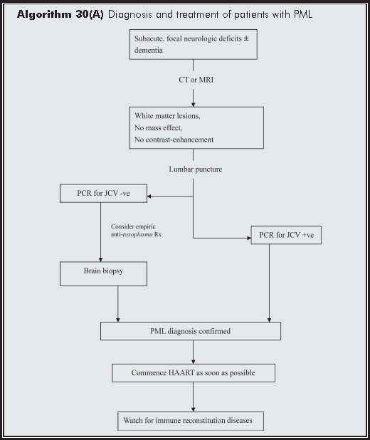
PML classically has a subacute clinical presentation with focal neurologic deficits, such as weakness, speech difficulties, unsteady gait and hemipareisis. Ophthalmic symptoms are relatively common, occurring as homonymous hemianopia which progresses to cortical blindness. These represent involvement of the white matter of the occipitoparietal lobe and the cerebellum. Seizure and headache are uncommon. Dementia manifesting as mental deficits in cognition, personality changes, and memory impairment are also common, but it is almost invariably associated with the focal neurologic deficits of PML. Radiologically, lesions are confined to the white matter, most commonly of the occipitoparietal lobe and without mass effect. MRI is superior to CT scan in the diagnosis of PML. The natural course of disease is one of inexorable progression to death in a median of 4 months, often punctuated by additional opportunistic infections.
In a patient with typical clinical features and especially a CD4 count <100/μL, the major differential diagnosis is limited to cerebral toxoplasmosis, primary CNS lymphoma or CMV encephalitis. CNS cryptococcomas and tuberculomas may occasionally cause diagnostic difficulties, especially if the appropriate CSF studies are not performed. Neuroimaging is very useful in diagnosis. In the CT scan, PML lesions are hypodense and exhibit no mass effect or contrast enhancement. In the more sensitive T2-wighted MRI, they are hyperintense, without gadolinium enhancement. White matter involvement is belied by the scalloped appearance of lesions. While they are most commonly found in the occipitoparietal lobes, other parts of the brain may also be affected, especially in combination with occipitoparietal involvement. These include basal ganglia, external capsule, the cerebellum and brainstem. In contrast to PML, toxoplasmosis lesions are usually ring-enhancing and CMV ventriculo-encephalitis is typically periventricular. In the latter, the gray matter is not spared and the clinical course rapidly progressive.
Routine tests of CSF are unremarkable in PML. The CSF pressure is normal. Protein may be high but glucose is normal. PCR for JC virus is relatively specific for PML but sensitivity is inadequate and very dependent on the primers employed. Sensitivity of this test has actually decreased in recent years, which is probably related to the widespread use of HAART.7 CSF should also be examined for Mycobacterium tuberculosis and Cryptococcus. If lymphoma or CMV encephalitis is a possibility, PCR for EBV and CMV respectively should be considered.
A working diagnosis of PML is generally based on the clinical presentation supported by the radiographic picture or a positive PCR test for JCV in the CSF. Definitive diagnosis requires a brain biopsy. With the use of HAART, PML may progress or present differently. An inflammatory component with cerebral oedema and radiographic enhancement by contrast may be apparent. As with other forms of immune reconstitution syndrome, PML may not manifest until after the start of HAART and signs of immune recovery or significant viral suppression are present.
There is no specific curative therapy for PML. Certain nucleosides, such as cytosine arabinoside (ara-C, cytarabine) and adenine arabinoside (ara-A, vidarabine), have been tried but with poor results. Cidofovir has in vitro activity against polyomavirus. Despite promising anecdotal reports, however, the drug failed to show benefit over the use of HAART in retrospective studies8 and a clinical trial.9 Other therapies that have been tried and failed include heparan sulfate, and interferon-α and -β.
As with other 'untreatable' complications of HIV such as Mycobacterium avium complex, microsporidiosis and CMV disease, use of HAART has been associated with improved prognosis of PML. Prolonged survival in patients on potent antiretrovirals was reported as a novelty in the early HAART era.10,11 These reports were later followed by systematic analyses which documented a significantly improved prognosis in the era of HAART. In Spain, 75 out of 111 (63.6%) patients with PML who received HAART survived a median of 2 years after the diagnosis.12 In France, there was a 63% reduction in risk of death in those who were on protease inhibitor-based HAART.13 In a large observational cohort in the US, the median survival time after diagnosis of PML was only 1 month. However, the 6-month survival time has increased from 10% for those without antiretroviral treatment to 68% for those given protease inhibitor-based HAART.14 These and other studies have also established the following prognostic factors of survival: receipt of HAART, high CD4 count at time of diagnosis, increase of CD4 count by >100/μL, low HIV viral load, PML as initial ADI, low-level or clearance of JCV in CSF, and lack of neurologic progression 2 months after diagnosis.15
Although the overall survival of PML has improved with the use of HAART, immune recovery has also been associated with an inflammatory reaction characterised by perivascular infiltration with lympho-plasmacytic cells. Radiographically, this is manifested as contrast enhancement and mass effect secondary to cerebral oedema; clinically, paradoxical worsening of symptoms is the predominant feature. Although such reactions are generally temporary, they may be so severe as to be fatal.16
Diagnosis of immune reconstitution disease (IRD) can be difficult. In every instance, other opportunistic infections should be ruled out. In fact, the very diagnosis of PML may have to be reconsidered, unless a definitive brain biopsy has been performed. Of note, the radiologic features of inflammation may lag behind the clinical worsening, further adding to diagnostic difficulty.17 A 'typical' IRD is characterised by unexpected clinical course or features, associated drop in viral load, rise in CD4 count, development of JCV-specific CTL, and a temporal relationship with the initiation of HAART. Although most forms of IRD are paradoxical 'exacerbation' of previously diagnosed PML, some are newly diagnosed cases that follow HAART-induced immune recovery. Systemic steroid is indicated for immediate management of severe reactions. Depending on circumstances, empiric treatment may have to be given for possible toxoplasmosis if that has not been excluded. Withdrawal of HAART is exceptional and should only be done in consultation with an expert in HIV care.
Recently, a form of HIV-associated leukoencephalopathy has also been described. It is characterised by very high levels of HIV RNA in the CNS and is usually associated with failure of HAART. A brain biopsy may be needed to differentiate from PML. Aetiology is unclear.18
Shankar SK, Satishchandra P, Mahadevan A, et al. Low prevalence of progressive multifocal leukoencephalopathy in India and Africa: is there a biological explanation? J Neurovirol. 2003;9 Suppl 1:59-67.
Dubois V, Moret H, Lafon ME, et al. JC virus genotypes in France: molecular epidemiology and potential significance for progressive multifocal leukoencephalopathy. J Infect Dis 2001;183:213-7.
Koralnik IJ, Wuthrich C, Dang X, et al. JC virus granule cell neuronopathy: A novel clinical syndrome distinct from progressive multifocal leukoencephalopathy. Ann Neurol 2005;57:576-80.
Du Pasquier RA, Corey S, Margolin DH, et al. Productive infection of cerebellar granule cell neurons by JC virus in an HIV+ individual. Neurology 2003;61:775-82.
Gasnault J, Kahraman M, de Goer de Herve MG, Durali D, Delfraissy JF, Taoufik Y. Critical role of JC virus-specific CD4 T-cell responses in preventing progressive multifocal leukoencephalopathy. AIDS 2003;17:1443-9.
Du Pasquier RA, Clark KW, Smith PS, et al. JCV-specific cellular immune response correlates with a favorable clinical outcome in HIV-infected individuals with progressive multifocal leukoencephalopathy. J Neurovirol 2001;7:318-22.
Marzocchetti A, Di Giambenedetto S, Cingolani A, Ammassari A, Cauda R, De Luca A. Reduced rate of diagnostic positive detection of JC virus DNA in cerebrospinal fluid in cases of suspected progressive multifocal leukoencephalopathy in the era of potent antiretroviral therapy. J Clin Microbiol 2005;43:4175-7.
Wyen C, Hoffmann C, Schmeisser N, et al. Progressive multifocal leukencephalopathy in patients on highly active antiretroviral therapy: survival and risk factors of death. J Acquir Immune Defic Syndr 2004;37:1263-8.
Marra CM, Rajicic N, Barker DE, et al. A pilot study of cidofovir for progressive multifocal leukoencephalopathy in AIDS. AIDS 2002;16:1791-7.
Elliot B, Aromin I, Gold R, Flanigan T, Mileno M. 2.5 year remission of AIDS-associated progressive multifocal leukoencephalopathy with combined antiretroviral therapy. Lancet 1997;349:850.
Power C, Nath A, Aoki FY, Bigio MD. Remission of progressive multifocal leukoencephalopathy following splenectomy and antiretroviral therapy in a patient with HIV infection. N Engl J Med 1997;336:661-2.
Berenguer J, Miralles P, Arrizabalaga J, et al. Clinical course and prognostic factors of progressive multifocal leukoencephalopathy in patients treated with highly active antiretroviral therapy. Clin Infect Dis 2003;36:1047-52.
Tassie JM, Gasnault J, Bentata M, et al. Survival improvement of AIDS-related progressive multifocal leukoencephalopathy in the era of protease inhibitors. Clinical Epidemiology Group. French Hospital Database on HIV. AIDS 1999;13:1881-7.
Dworkin MS, Wan PC, Hanson DL, Jones JL. Progressive multifocal leukoencephalopathy: improved survival of human immunodeficiency virus-infected patients in the protease inhibitor era. J Infect Dis 1999;180:621-5.
Skiest DJ. Focal neurological disease in patients with acquired immunodeficiency syndrome. Clin Infect Dis 2002;34:103-15.
Safdar A, Rubocki RJ, Horvath JA, Narayan KK, Waldron RL. Fatal immune restoration disease in human immunodeficiency virus type 1-infected patients with progressive multifocal leukoencephalopathy: impact of antiretroviral therapy-associated immune reconstitution. Clin Infect Dis 2002;35:1250-7.
Silva MT, Pacheco MC Jr, Vaz B. Inflammatory progressive multifocal leukoencephalopathy after antiretroviral treatment. AIDS 2006;20:469-71.
Langford TD, Letendre SL, Marcotte TD, et al. Severe, demyelinating leuko-encephalopathy in AIDS patients on antiretroviral therapy. AIDS 2002;16:1019-29.