Owen TY TSANG
Toxoplasma gondii (T. gondii) is an obligate intracellular protozoan of worldwide distribution. Its infection in immunocompetent individuals has generally been regarded as being of little significance, since an effective cell-mediated immunity towards T. gondii results in control though not an eradication of the infection. Latent infection is characterised by the persistence of the parasite in the host's tissue. A defect in cell-mediated immunity of the infected person predisposes to the risk of reactivation of the organism. The commonest clinical manifestation in HIV-infected patients is encephalitis.
T. gondii exists in three functionally distinct forms: oocysts, tachyzoites and tissue cyst containing bradyzoites. Oocyst is the infective form of T. gondii and is shed by the definitive host, the cat. It contains sporozoites which, upon ingestion by mammals (including human), develop into tachyzoites and enter all nucleated host cells. Tachyzoites are the invasive form of the organism found in acute infection or reactivation. They multiply rapidly, lead to cell rupture and invasion of nearby cells, or are transported to other parts of the body via the blood and lymphatic system. They are transformed into tissue cysts under the pressure of the inflammatory response. Tissue cyst is the dominant form of the parasite containing hundreds or thousands of bradyzoites. They can be infective after ingestion by the intermediate or definitive hosts. The commonest sites of cyst formation are the brain, skeletal and cardiac muscles. Bradyzoite can be released from the cyst and transformed back into tachyzoites in immunocompromised host.
The seroprevalence of T. gondii increases with age and varies with population group and geographic location - which can be over 50% in Europe and other tropical countries.1 The prevalence of T. gondii in a group of randomly selected inhabitants in Hong Kong was 9.8%.2
In the United States, about one-third of HIV-infected patients have antibodies against T. gondii.3 Previous studies showed that toxoplasmic encephalitis (TE) ultimately develop in 25-50% of patients with AIDS, especially those with CD4 T-cell counts less than 100/μL.4 Most of the encephalitic cases result from reactivation of a latent infection. However, with the introduction of potent antiretroviral therapy and primary prophylaxis, the risk of toxoplasmosis has declined significantly. In Hong Kong, toxoplasmosis accounts for about 2% of all AIDS-defining illnesses.5
Toxoplasmosis in HIV-infected patients manifests primarily as encephalitis although a minority of them could also present as chorioretinitis, pneumonitis or disseminated infection. Clinical presentation of TE depends on the size, number and location of the brain lesions, and the immune status of the host. It is usually subacute in onset with focal neurologic signs frequently accompanied by fever, altered mental state and headache. Cerebellar, subcortical or cortical lesions can be present in over 50% of the infected cases, resulting in hemipareisis, ambulatory, gait or speech abnormalities.6 A significant proportion of encephalitic patients can also present with neuropsychiatric disorders including psychosis, dementia, anxiety and personality disorder.7
The differential diagnoses of toxoplasmosis in an HIV patient are:
(a) Lymphoma of the central nervous system (CNS)
(b) Progressive multifocal leukoencephalopathy
(c) Tuberculosis, including tuberculoma
(d) Focal CNS lesions caused by other infection including Cryptococcus neoformans, Aspergillus spp, Mycobacterium tuberculosis, and Nocardia spp
(e) Cytomegalovirus or Herpes simplex encephalitis
(f) Bacterial brain abscess
Diagnosis of TE is made by clinical, serologic, radiological, histological or molecular methods, or by a combination of these. Clinical signs of CNS toxoplasmosis are often non-specific. However, an HIV-infected patient with CD4 T-cell counts less than 100/μL presenting with compatible focal CNS signs should alert clinician of the diagnosis.
Anti-T. gondii IgG antibodies start to rise 1 to 2 weeks after acquisition of the infection and peak at 6 to 8 weeks. They decline gradually over the next 1 to 2 years, but they can persist for life in some cases.8 Almost all HIV-infected patients with TE have IgG antibodies & thus the absence of them make the diagnosis of toxoplasmosis unlikely. However, a measurement of IgG level could not differentiate between recently acquired and distant infection. Anti-T gondii IgM antibodies tend to appear earlier and decline faster than IgG antibodies. The absence of IgM virtually excludes recent infection in immunocompetent patients. However, they can remain elevated for years and thus are not recommended for routine use.
Magnetic resonance imaging (MRI) is more sensitive than Computed tomography (CT) scan in diagnosing toxoplasmosis from encephalitic brain lesions. Typical radiological findings comprise of bilateral, multiple, ring-enhancing lesions over basal ganglia and corticomedullary junctions of cerebral hemisphere.9 Surrounding oedema and mass effect are present in varying degrees. The most important differential diagnosis to consider for brain lesion in an AIDS patient is CNS lymphoma. Features that favour the diagnosis of T. gondii encephalitis over CNS lymphoma include: subcortical lesions, more than 3 lesions, absence of ependymal or leptomeningeal involvement, marked peri-lesional oedema, absence of hyperattenuation on nonenhanced CT scans or slender uniform ring-enhancing foci.10 Increased uptake in newer imaging techniques like single-photo emission computed tomography (SPECT) or positron emission tomography (PET) can enhance the specificity for the detection of CNS lymphoma.
Brain biopsy showing tachyzoites or cyst provides a definitive diagnosis for TE. However, it is rarely considered nowadays since the empirical therapy of suspected toxoplasmosis can usually confirm the diagnosis. Biopsy is indicated in patients who fail to respond to anti-T gondii therapy or are suspected to have diagnosis other than toxoplasmosis.
Cerebrospinal fluid and blood sample for toxoplasmic polymerase chain reaction (PCR) yields a poor and variable sensitivity11 and a positive PCR in brain tissue does not necessarily indicate active infection.
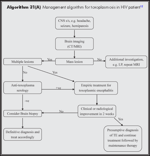
Since TE was the most common cause of focal CNS lesions in AIDS patients before the era of HAART (highly active antiretroviral therapy), empiric anti-T. gondii therapy used to be the standard approach. However, the incidence of TE in AIDS patients has significantly decreased since the introduction of HAART and the widespread use of prophylaxis. Therefore, this empiric therapeutic approach may miss or delay the appropriate work-up and management of important diagnosis like CNS lymphoma. Algoritum 31(A) illustrates the management approach to TE in HIV patient.12 In general, the presence of multiple brain lesions with CD4 T-cell counts of less than 100/μL and positive anti-T. gondii antibodies in an HIV patient who is not taking anti-T. gondii prophylaxis is highly predictive of TE.
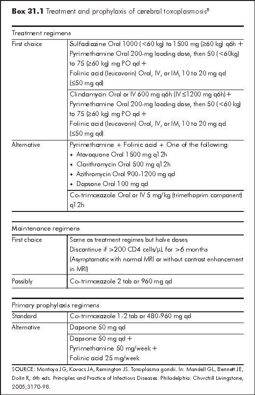
Combination of pyrimethamine/sulfadiazine and folinic acid is considered the standard regime for the treatment of TE (Box 31.1).8 Unfortunately sulfadiazine is not available in Hong Kong. Clindamycin can be used instead of sulfadiazine in this regard. Infected patient should be treated for at least 4-6 weeks after the resolution of all signs and symptoms. It is important to note that, as a result of the myelotoxicity of sulfonamides and pyrimethamine, "folinic acid" instead of "folic acid" should be used since the latter will reverse the action of pyrimethamine. The efficacy of Trimethoprim/sulfamethoxazole (co-trimoxazole) appears to be comparable to that of pyrimethamine/sulfadiazine in AIDS patients.13 Short course of corticosteroid can be used in TE patient with significant cerebral oedema and elevated intracranial pressure.
In one study 51% of patients with TE developed clinical response to anti-T. gondii therapy within the first 3 days and 91% by day 14.6 Other investigation including brain biopsy should be considered if there is no improvement by 2 weeks or when there is deterioration by day 3.6 Over 90% of patients have radiological response by 2 weeks of therapy. Monitoring brain CT/MRI every 4-6 weeks is suggested until there is complete resolution of the lesions.
After the acute treatment of TE, maintenance therapy (secondary prophylaxis) should follow since the current anti-T. gondii therapy cannot eradicate tissue cysts. Normally the same medications used in the acute phase could be continued at half dose to this effect (Box 31.1).
Primary prophylaxis should be considered in HIV patient with CD4 cell counts less than 100/μL. Use of co-trimoxazole for the prophylaxis of Pneumocystis jiroveci pneumonia (PCP) could also provide protection against toxoplasmosis. Other alternatives include high dose dapsone alone or dapsone plus pyrimethamine. Both primary or secondary prophylaxis can be discontinued when the patient's CD4 cell count has returned to over 200/μL for at least 6 months.14
HIV-infected persons should be tested for baseline IgG antibodies to Toxoplasma to detect latent infection with T. gondii. All HIV-infected persons should be counseled regarding exposure to toxoplasmic infection:15
(a) Avoid eating raw or undercooked meat, including undercooked mutton, beef, pork, or venison
(b) Wash hands after contact with raw meat, after gardening or other contact with soil
(c) Wash fruits and vegetables well before eating them raw
(d) Avoid handling cats' litter and wash hands thoroughly after changing the litter box
(e) Keep cats inside, and do not to adopt or handle stray cats
(f) Feed cats only with canned or dried commercial food or well-cooked table food, not raw or undercooked meats
1. Montoya JG, Kovacs JA, Remington JS. Toxoplasma gondii. In: Mandell GL, Bennett JE, Dolin R, 6th eds. Principles and Practice of Infectious Diseases. Philadelphia: Churchill Livingstone, 2005;3170-98.
2. Hunter CA & Reichmann G. Immunology of toxoplasma infection. In Joynson DHM, Wreghitt TG. Toxoplasmosis: a comprehensive clinical guide. Cambridge: Cambridge University Press, 2001;43-57.
3. Montoya JG, Liesenfeld O. Toxoplasmosis. Lancet 2004;363:1965-76.
Tenter AM, Heckeroth AR, Weiss LM. Toxoplasma gondii: from animals to humans. Int J Parasitol 2000;30:1217-58.
Ko RC, Wong FW, Todd D, Lam KC. Prevalence of Toxoplasma gondii antibodies in the Chinese population of Hong Kong. Trans R Soc Trop Med Hyg 1980;74:351-4.
Grant IH, Gold JW, Rosenblum M, Niedzwiecki D, Armstrong D. Toxoplasma gondii serology in HIV-infected patients: the development of central nervous system toxoplasmosis in AIDS. AIDS 1990;4:519-21.
Luft BJ, Remington JS. Toxoplasmic encephalitis in AIDS. Clin Infect Dis 1992;15:211-22.
Department of Health. Hong Kong STD/AIDS Update: a quarterly surveillance report. July 1998;4(3):7.
Luft BJ, Hafner R, Korzun AH, et al. Toxoplasmic encephalitis in patients with the acquired immunodeficiency syndrome. Members of the ACTG 077p/ANRS 009 Study Team. N Engl J Med 1993;329:995-1000.
Torrey EF, Yolken RH. Toxoplasma gondii and schizophrenia. Emerg Infect Dis 2003;9:1375-80.
Montoya JG, Kovacs JA, Remington JS. Toxoplasma gondii. In: Mandell GL, Bennett JE, Dolin R, 6th eds. Principles and Practice of Infectious Diseases. Philadelphia: Churchill Livingstone, 2005;3170-98.
Levy RM, Rosenbloom S, Perrett LV. Neuroradiologic findings in AIDS: a review of 200 cases. AJR Am J Roentgenol 1986;147:977-83.
Dunn IJ, Palmer PE. Toxoplasmosis. Semin Roentgenol 1998;33:81-5.
Parmley SF, Goebel FD, Remington JS. Detection of Toxoplasma gondii in cerebrospinal fluid from AIDS patients by polymerase chain reaction. J Clin Microbiol 1992;30:3000-2.
Wong KH. Toxoplasmosis. In: Chan K, Wong KH, Lee SS. HIV manual 2001. Red Ribbon Centre 2002;193-8.
Torre D, Casari S, Speranza F, et al. Randomized trial of trimethoprim-sulfamethoxazole versus pyrimethamine-sulfadiazine for therapy of toxoplasmic encephalitis in patients with AIDS. Italian Collaborative Study Group. Antimicrob Agents Chemother 1998;42:1346-9.
Mofenson LM, Oleske J, Serchuck L, et al. Treating opportunistic infections among HIV-exposed and infected children: recommendations from CDC, the National Institutes of Health, and the Infectious Diseases Society of America. MMWR Recomm Rep 2004;53(RR-14):1-92.
1999 USPHS/IDSA guidelines for the prevention of opportunistic infections in persons infected with human immunodeficiency virus. U.S. Public Health Service (USPHS) and Infectious Diseases Society of America (IDSA). MMWR Recomm Rep 1999;48(RR-10):1-59, 61-6.